Tracking Vesicle Dynamics at the Spicule Tip
Why I started this experiment
One question that interests me is how mineral-bearing vesicles behave near the spicule growth zone during skeletogenesis.
Are vesicles diffusing randomly?
Are they guided toward the biomineralization compartment?
Can actomyosin dynamics influence their motion?
These questions motivated this imaging experiment.
Experimental setup
Embryos were imaged using spinning disk confocal microscopy.
Markers used:
- Calcein to visualize calcium-containing vesicles
Focus was placed near the spicule tip.
Representative image
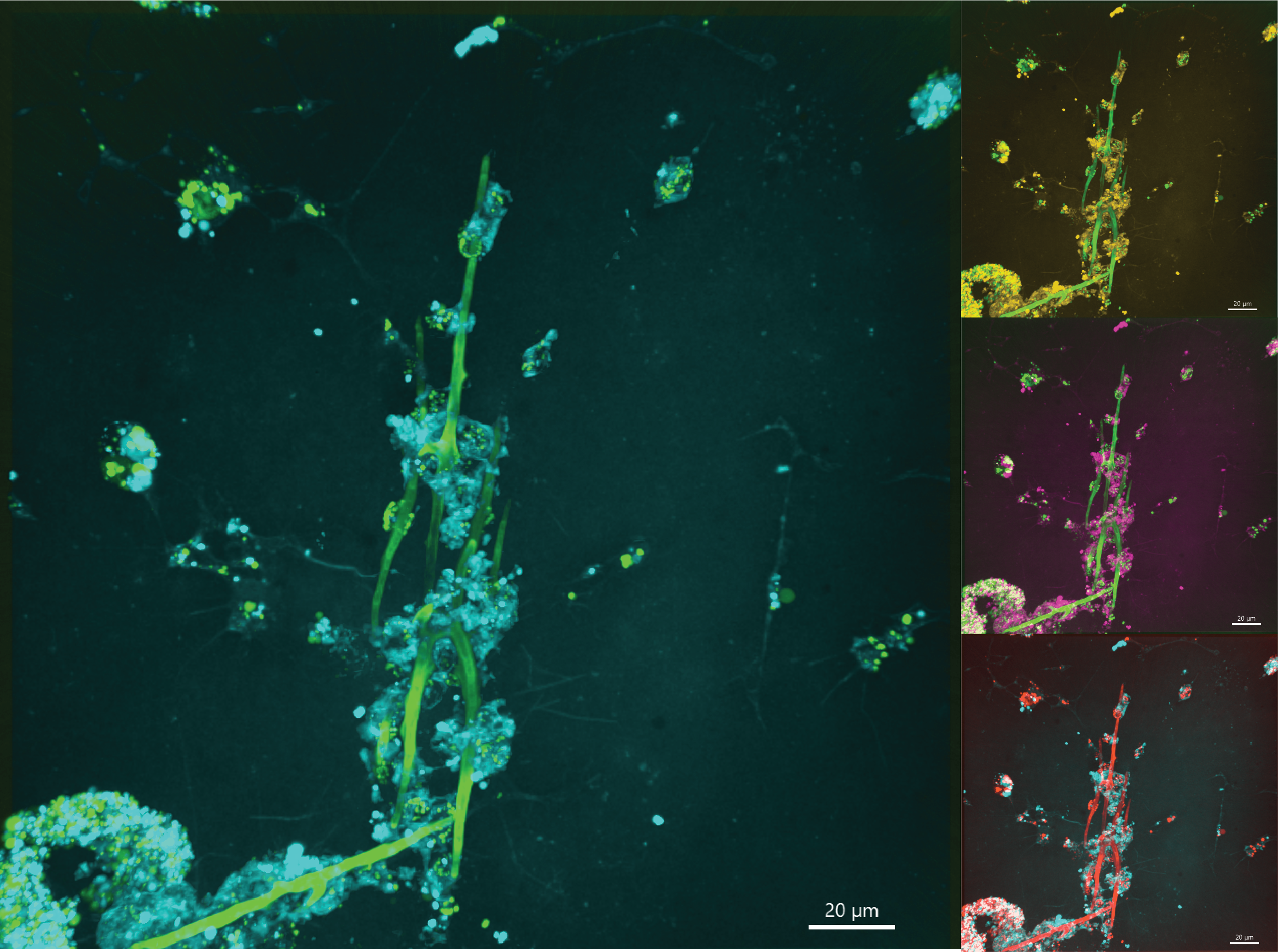
This image shows the region of interest near the spicule growth zone used for live imaging analysis.
Initial observations
Some preliminary patterns appeared interesting:
- Vesicles appear enriched near the growth region
- Motion seems partly diffusive, though not purely random
- Some trajectories may suggest local regulation
These are early observations and require quantitative analysis.
Questions I am exploring
Current questions:
-
What is vesicle diffusion behavior near the tip?
-
Does actomyosin contractility influence vesicle mobility?
-
Is there evidence for directed trafficking versus active diffusion?
Next steps
Planned analyses:
- Track vesicle trajectories
- Quantify diffusion coefficients
- Measure directionality index
- Compare control vs perturbation conditions
This notebook entry documents early thoughts from an ongoing project.